



- Technical Specifications
- Description
| Brand | SonoScape |
|---|---|
| Model | P40 Elite |
| Product Type | AI ultrasound system |
| Software Tools | S-Fetus and Auto OB Plus |
| Clinical Focus | Obstetrics and gynaecology |
Description
SonoScape P40 Elite – Full OB/GYN AI Package (AI Time-Saving Ultrasound )
P40 Elite is a high-performance ultrasound system designed to make OB/GYN exams faster and more consistent.
AI tools help automate measurements and reduce manual steps, so doctors can scan and report fatser.
AI / AUTOMATION
- S-Fetus 5.0 S-Fetus (Advanced) automatic acquisition of key fetal scan planes and measurements for faster, more standardized exams.
- Auto OB one-click fetal biometry package (reduces user-dependent variability and speeds up routine measurements).
- Auto measurements
- Auto Trace calculations
AI inOB/GYN
- S-GYN
- S-Follicle
- S-Uterus
- Auto Face
IMPROVED IMAGE QUALITY AND ADVANCED FLOW TECHNOLOGY
- Micro F Detects extremely small and slow blood flow in microvessels and display their perfusion with high detail , Micro F allows you to visualize microvascular flow that is often not visible with conventional Doppler techniques
- SR Flow Improves spatial resolution of flow Shows vessels more clearly and continuously , SR Flow improves both sensitivity and resolution, so you can see slow blood flow more clearly and with better vessel definition compared to conventional Doppler .
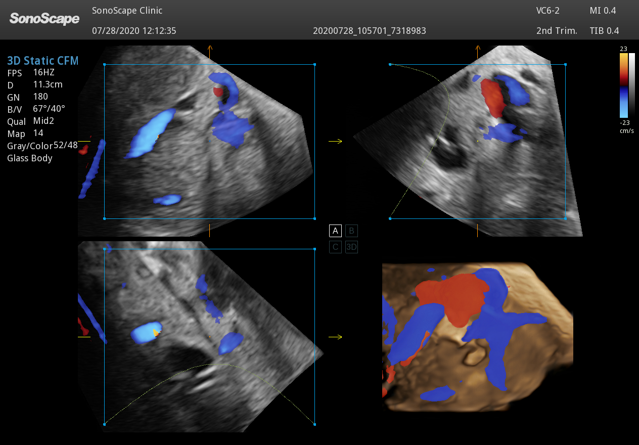
- BRIGHT FLOW 3D-like visualization of blood flow.
- Better visual understanding of vessels , Works in CFM / PDI / Micro F / SR Flow modes
Automatically adjusts 2D flow visualization
Creates a stereo 3D image without the need of using volume probe
- Better visual understanding of vessels , Works in CFM / PDI / Micro F / SR Flow modes
- HQ Scan nand μ-Scan with Speckle noise reduction.
- Smoother image
- Less grain
- Sharper structures
- Compound imaging : Combines images from multiple angles.
- Reduces artifacts
- Improves tissue clarity
- HPRF high pulse repetition frequency doppler It is a special Doppler mode that allows the system to measure higher blood flow velocities than standard PW Doppler.
BASIC FEATURES : B mode , M mode , CFM (color flow mapping for flow direction) , PDI & DPDI (shows how blood flows , can detect slow flow as well )
BENEFITS OF P40 ELITE :
- Cleaner images, less re-scanning thanks to advanced processing and speckle reduction tools.
- Stronger patient communication with wireless sharing of images/clips and simple reporting transfer options.
- OB/GYN power package for standard planes, biometry, 3D/4D rendering, and volume analysis.
OB/GYN AI – Saves Time Automated standard planes + biometry
- Auto OB Plus
- Auto NT semi-automatic nuchal translucency measurement to support standardized 1st trimester workflow.
Smarter gynecology & fertility workflow
- AVC Follicle automatic follicle counting + volume calculation from volume data (excellent for IVF/infertility follow-up).
- S-Follicle – automatic follicle recognition and size measurement.
- S-Endo – automatic endometrium recognition and thickness measurement.
- S-Ovary / S-Uterus – automated 2D measurements and annotation support for faster reporting.
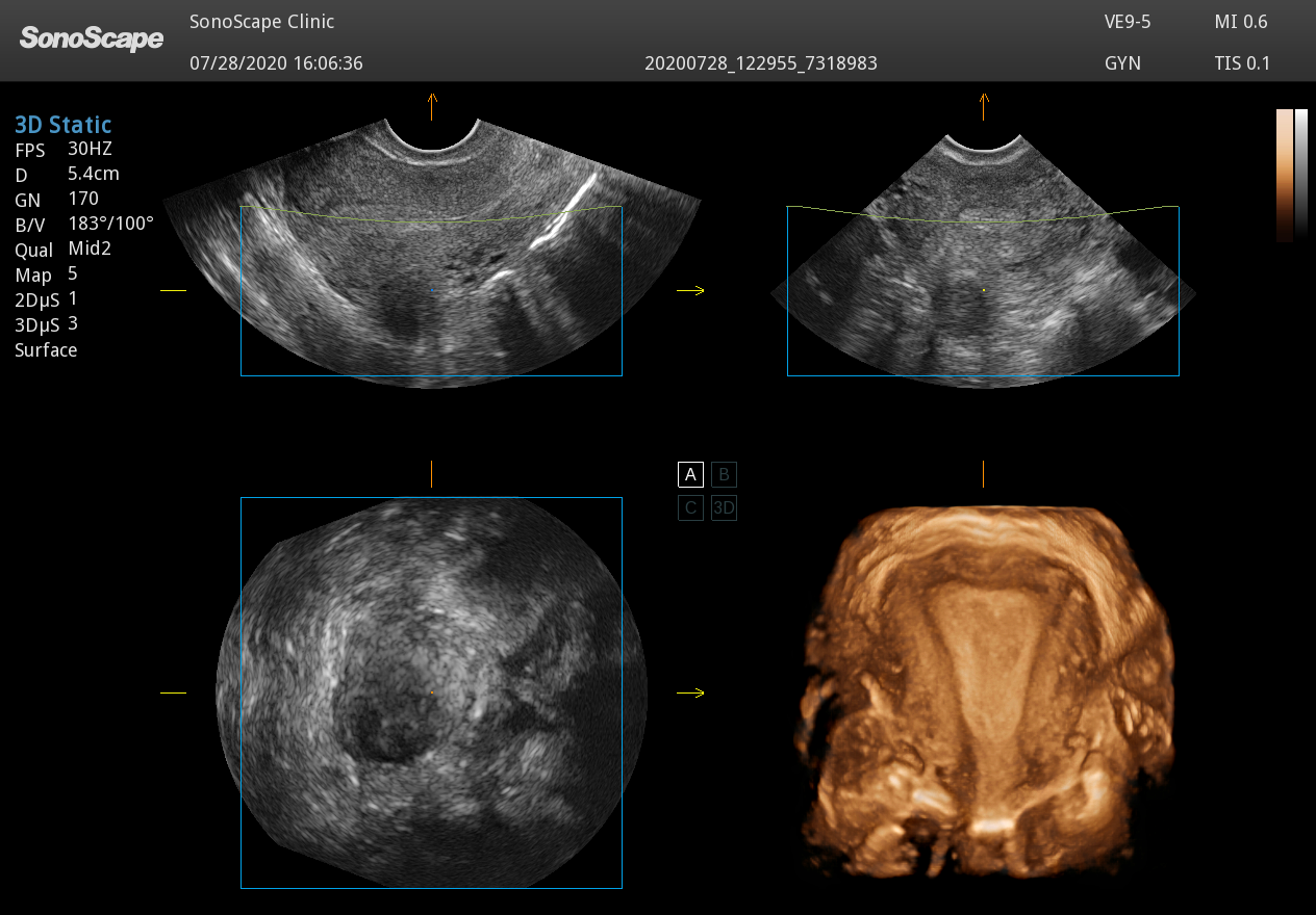
- Auto-Uterine (3D) – automated 3D uterine imaging (requires 3D/4D capability).
- S-PF (2D)
AI-enhanced 3D/4D + fetal assessment tools with the volumetric probes: VC6-2 (volume convex) & VE9-5 (volume endocavity)
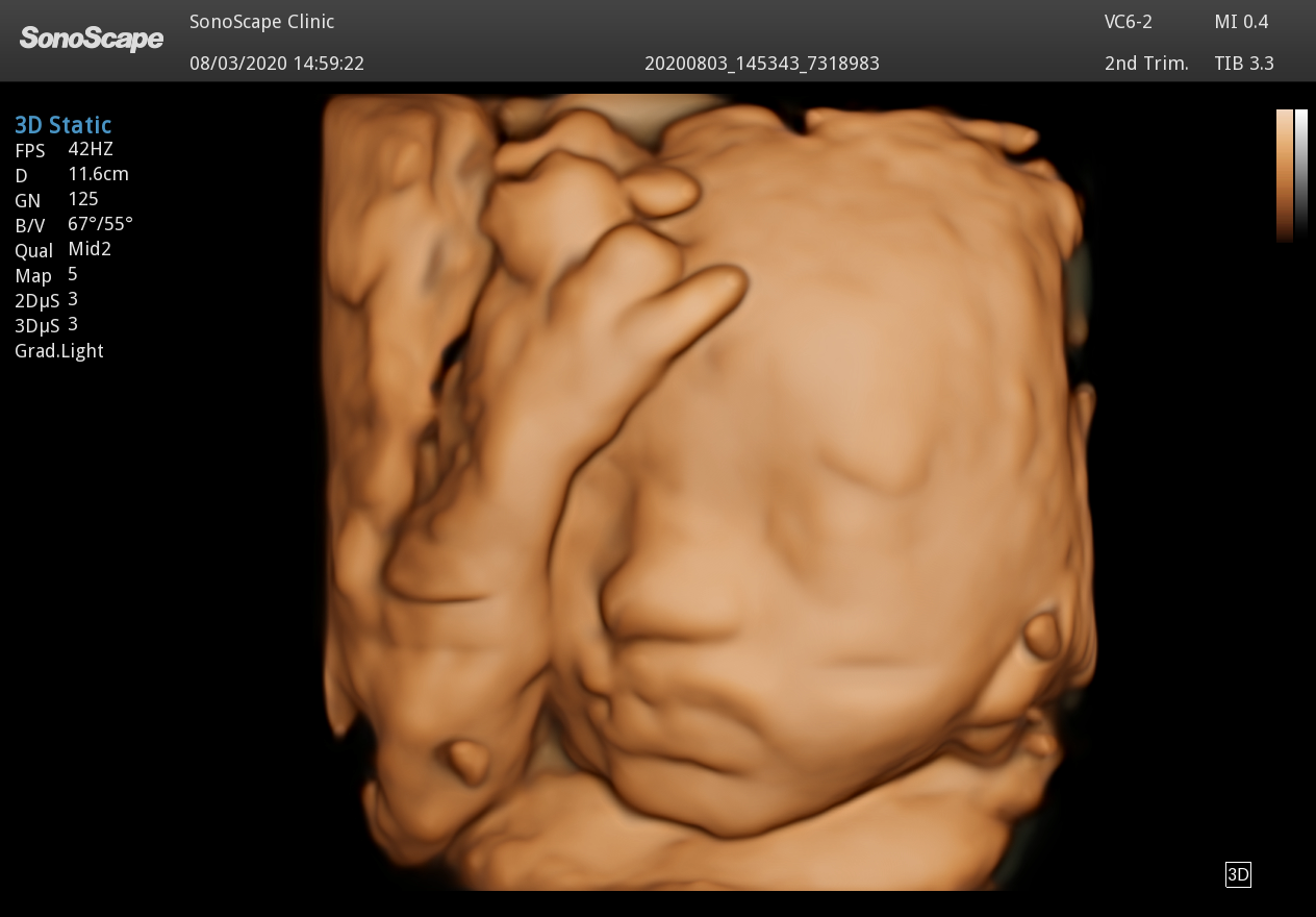
- Auto Face (3D) – intelligent fetal facial detection and optimization (less time fighting shadowing/occlusions).
- S-Spine – intelligent fetal spine identification with automatic 3D imaging support.
- S-Hip – one-button recognition of hip joint structure (fast screening support).
- STIC – spatio-temporal imaging correlation for fetal heart volume acquisition (3D/4D module required).
- VOCAL (3D Volume Calculation): Semi-automated 3D volume measurement for lesions and masses. Trace the structure on rotation planes and the system calculates accurate volume for follow-up and reporting.
VCI (Volume Contrast Imaging): Cleaner, low-noise volume slices with adjustable thickness (2–20 mm) for clearer borders and faster interpretation.
FreeVue (Any-Plane Coronal View): FreeVue lets you create the coronal view from a 3D volume by cutting the uterus/lesion any way you want (straight line, curve, trace, polyline). So instead of fighting for the perfect 2D plane, you acquire one good volume and then reconstruct the exact slice you need. - S-Live / S-Live Silhouette / S-Depth – realistic rendering modes for clearer anatomy visualization in complex cases.
How This Improves the Doctor’s Day
- Fewer clicks per exam: automate the repetitive steps (planes, measurements, optimization), so you stay focused on interpretation.
- More consistent results: AI-driven measurement support reduces operator variability especially across busy teams or multi-doctor practices.
- Shorter exam times: faster biometry + auto tools = higher throughput without sacrificing documentation quality.
- Cleaner reporting workflow: built-in connectivity and report export options reduce end-of-day admin load.
- Better patient communication: easy sharing tools help patients understand findings without slowing you down.
Imaging Performance & Clinical Tools
- Full Doppler suite: Color, PW, CW, Power Doppler, Directional Power Doppler, Triplex.
- High-detail imaging: Tissue Harmonics, Spatial Compound, Tissue Specific Imaging.
- μ-Scan (2D/3D) speckle reduction for clearer tissue detail and border definition.
- HQ-Scan contrast enhancement for improved visualization.
- SR Flow + Micro F for improved display of slow flow and microvascular structures.
- Bright Flow for more intuitive 3D-style visualization of blood flow.
- C-xlasto elastography (strain elastography with strain ratio) depending on probe/configuration. Workflow, Training & Connectivity
- Sono-Help – built-in scanning tutorials (probe placement, anatomy, standard image examples) for faster staff training.
- Sono Assistant – customizable workflow protocols for vascular and obstetrics applications (standardize exams, reduce keystrokes).
- Sono-Drop – Sono-drop provides a fast and convenient ultrasound image transmission between P40 ELITE and the patients’ smart devices. The bond between clinicians and patients are supposed to be strengthened through more frequent communication.
- Sono-Synch –Real-time interface and camera sharing, enabled by Sono-synch, makes it possible to connect two ultrasound in a remote distance and perform remote medical consultation and tutorial.
- Sono-Trans – export PDF reports, images and cine loops to a shared Windows folder.
- DICOM 3.0 – Store, Worklist, MPPS, Print, Structured Reporting, etc.
- Built-in DVR – record screen + audio for teaching, documentation, and case review.
Configuration
- P40 Elite trolley system with 21.5″ High Resolution Medical Monitor + Multi-directional articulating monitor arm + 13.3” touch screen
- 2TB storage
- 4 active probe ports + parking port (plus dedicated pedoff port)
- Recomended convex probe : C1-6
- Available Volumetric probes: VC6-2 (volume convex) & VE9-5 (volume endocavity)
- Full OB/GYN AI Package (some modules require 3D/4D probes )
Note: Availability of certain advanced modules depends on selected probes and 3D/4D configuration.
Ideal For
- OB/GYN clinics aiming for faster exams + standardized reporting
- IVF/infertility centers needing follicle/volume tools
- Hospitals and imaging centers that want strong connectivity + training support
Request a Quote / Demo call us on 22441043 to arrange a meeting for presentation.
Learn more about sonoscape here !

μ-Scan+
A new generation μ-Scan+, available for both B and 3D/4D modes, is more delicately engineered to distinguish tissue and artifacts. In the meantime of reducing speckles, it can improve image uniformity and enhance border continuity to provide authentic presentation of details and enhanced lesion display.

SR-Flow The separation of blood flow and tissue signal becomes more easily with SR-Flow given the use of a highly effective filter technology. It enables a dynamic and vivid Doppler display with high sensitivity while ensuring a realistic evidence for detection of slow flows.

Micro F provides an innovative method to expand the range of visible flow in ultrasound, especially for visualizing hemodynamic for tiny vessels. Detailed views of blood flow in relation to nearby tissue also render more diagnostic confidence to evaluate lesions and tumors.
Intelligent Solution at Your Fingertips
Routine over-repetitive exams and complicated operation are stressing out ultrasound clinicians. Intelligent solution provided by ELITE streamlines parts of the workflow to improve remarkably efficiency, with AI-powered tools including measurement, parameter adjustments, image optimization, etc.

S-Fetus
Based on a big data dependable deep learning algorithm, S-Fetus is a brilliant one-stop solution for automatic standard plane acquisition and measurement. With just one click, common fetal biometry results are obtained with high intelligence, accuracy and efficiency, aiming for an unprecedented ease during operation.

Auto OB Plus
Fast and highly efficient fetal biometry is achieved by the help of Auto OB. Meanwhile, more consistent results given by this deep learning based method can effectively reduce user-dependent variability.

Auto NT
Auto NT provides semi-automatic, standardized measurements of the nuchal translucency thickness in 2D image and reduces operator dependency on the results.

Auto Bladder
One key bladder wall tracing and volume measurement from Auto Bladder can efficiently provide more accurate contour and results, which is not subject to the bladder shape and size.

Auto IMT
Auto IMT makes the measurement of anterior and posterior intima-media thickness much easier with simple placement of the ROI.

AVC Follicle
High efficiency of follicle analysis is achieved by AVC Follicle, a volume-data based automatic follicular calculation including the number and volume. Follicles are sorted by sizes in the results and rendered in different colors for better visualization.
Fast and Efficient Optimization

Auto B/C
Imaging parameter adjustment is now no more done in a laborious manner. Auto B/C helps to optimize the image quality under B and color Doppler mode within just one click. Multiple parameters such as gain, TGC, ROI position, steering angle, etc., are all included in this full automation method.

Auto Face
3D fetal face visualization is significant for face anomalies diagnosis. The removal of occlusions and artifacts, such as cord, placenta, uterus and extremities, can be simply accomplished by Auto Face to get an optimal view of fetal face.
U

MFI Time
MFI Time provides a color coded parametric view, indicating the uptake time of contrast agents in different perfusion phases to better differentiate tissues.

S-Live Silhouette
Through the application of an artificial light source and shadowing effect, S-Live Silhouette sees through the surface and clearly delineates the outlines of bone, organs, cavities, vessel walls and other internal structures. It is a beneficial tool for identifying normal anatomy and diagnosing complex congenital malformations.

Color 3D
Available on color and power Doppler mode, Color 3D applies advanced rendering to blood flow to produce more intuitive and natural hemodynamics of vascular networks with speed and direction information, especially for umbilical cords.

Stress Echo
A straightforward template for clinicians to take multiple dynamic images at rest and after stress and make side by side comparison. Professional wall motion bulls-eye scoring and reporting is provided for further effective evaluation of cardiac muscle viability.

Myocardium Quantitative Analysis (MQA)
Precise quantitative measurement on myocardial mechanics is achieved by MQA based on real-time sensitive wall motion tracking. It provides global and regional assessment including strain, strain rate, displacement, velocity, etc.
Recomnded probes
Sonoscape Systems in Cyprus call for demostration on 22441043